Introduction
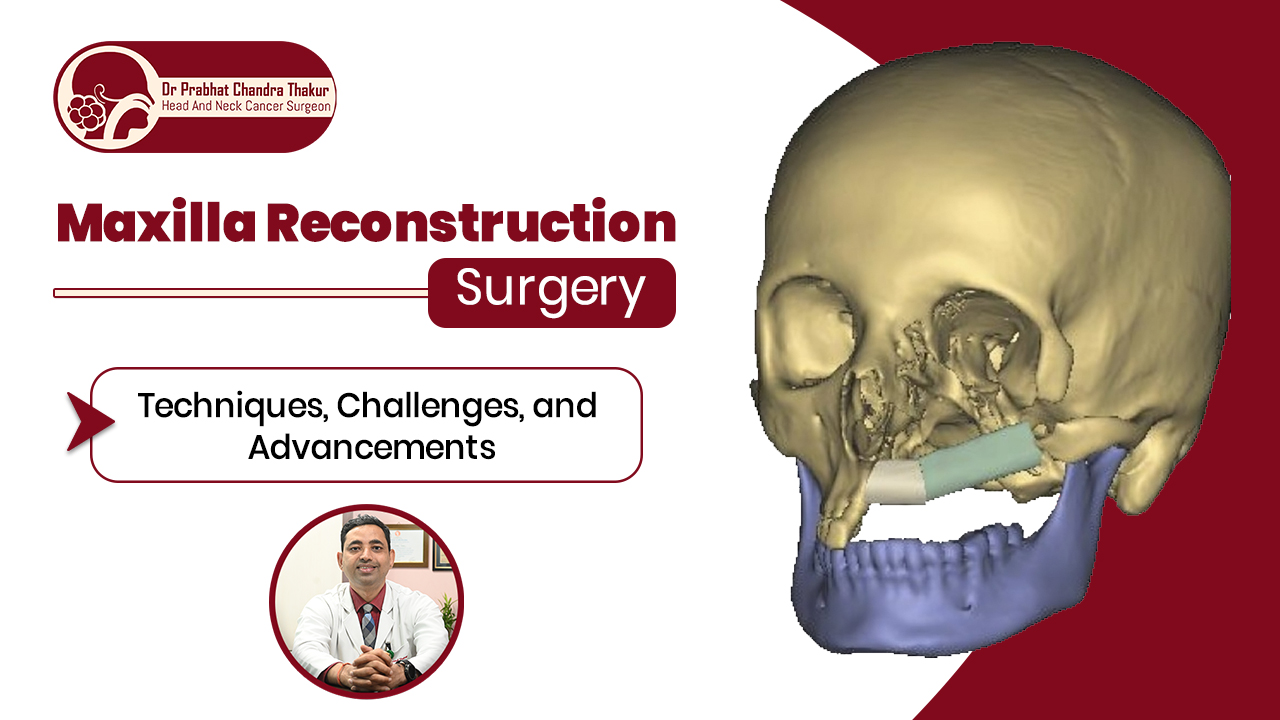
Maxilla reconstruction surgery is an evolving field in maxillofacial surgery, aimed at restoring both function and aesthetics for patients with maxillary defects. These defects may arise due to trauma, cancer resections, congenital anomalies, or infections, leading to impaired speech, swallowing, mastication, and facial deformity. Unlike mandibular reconstruction, maxillary reconstruction presents unique challenges due to its complex anatomical structure and functional demands.
Dr. Prabhat Chandra Thakur, one of Nepal’s leading head & neck surgeons, specializes in advanced reconstructive techniques to restore maxillary defects. His expertise in microsurgical reconstruction, free flaps, and maxillofacial surgery has made him a trusted name in Nepal for patients seeking maxilla reconstruction surgery.
Why Maxilla Reconstruction Is Important?
A defect in the maxilla can significantly impact a patient’s quality of life. The maxillary bone provides:
- Structural support for the midface and orbit
- Separation between the oral and nasal cavities
- Foundation for mastication and speech
- Aesthetic integrity of the face
Maxillary defects can be rehabilitated through prosthetic obturators or surgical reconstruction, with surgical reconstruction offering more long-term benefits in function and aesthetics.
Common Causes of Maxillary Defects
- Head & Neck Cancer Surgery (Maxillectomy)
- Squamous cell carcinoma, sinus tumors, or other malignancies often require surgical removal of the maxilla.
- Squamous cell carcinoma, sinus tumors, or other malignancies often require surgical removal of the maxilla.
- Trauma & Accidents
- Road traffic accidents, gunshot wounds, or falls can cause severe maxillary injuries.
- Road traffic accidents, gunshot wounds, or falls can cause severe maxillary injuries.
- Congenital Defects
- Cleft palate and other craniofacial anomalies may require reconstruction for functional restoration.
- Cleft palate and other craniofacial anomalies may require reconstruction for functional restoration.
- Infections & Osteonecrosis
- Severe infections, such as osteomyelitis, can lead to bone loss requiring reconstruction.
Reconstructive Techniques for Maxillary Defects
1. Prosthetic Rehabilitation (Obturators)
Historically, maxillary defects were rehabilitated using obturator prostheses, which cover the palatal defect to aid in speech and swallowing. While obturators are simple and non-invasive, they often fail in larger defects due to:
- Poor stability
- Difficulty in retention
- Oro-nasal leakage issues
Recent studies suggest that free flap reconstruction provides better quality of life compared to obturators, particularly in cases of large or posteriorly placed defects.
2. Surgical Reconstruction
Surgical reconstruction remains the gold standard for moderate to severe maxillary defects. Several reconstructive methods exist:
A. Regional Soft Tissue Flaps
- Temporalis Myofascial Flap – Offers soft tissue coverage but lacks structural support.
- Buccal Fat Pad Flap – Suitable for small lateral defects.
- Facial Artery Myomucosal Flap (FAMM) – Used for limited defects with good results.
B. Free Tissue Transfer (Free Flaps)
Free flap techniques provide bone support, orbital integrity, and dental rehabilitation. The most commonly used free flaps include:
- Free Fibula Flap – Best for alveolar and orbital support; allows for dental implants.
- Deep Circumflex Iliac Artery (DCIA) Flap – Provides a good contour for the midface but has a shorter vascular pedicle.
- Scapular Free Flap – Offers excellent soft tissue coverage with a reliable blood supply.
- Radial Forearm Free Flap – Useful for soft tissue coverage, but lacks bone support.
3. Use of Titanium Mesh & Alloplastic Implants
For orbital support, titanium mesh is sometimes used in combination with soft tissue or bone grafts. However, mesh extrusion rates are high, especially after post-operative radiation therapy.
Classification of Maxillary Defects
Multiple classification systems exist for maxillary defects, but Brown’s Classification is widely used. It divides defects into:
- Class I – Palatal defects (small, moderate)
- Class II – Alveolar defects with some palatal involvement
- Class III – Defects involving orbital floor support
- Class IV – Extensive defects including orbital exenteration
Dr. Prabhat Chandra Thakur follows an individualized approach based on defect severity, patient health, and functional goals to choose the best reconstructive option.
Challenges in Maxillary Reconstruction
Maxillary reconstruction presents multiple challenges, including:
- Oncological Safety – Ensuring tumor clearance while preserving function.
- Orbital Support – Preventing enophthalmos (sunken eye) and maintaining normal eye position.
- Dental Rehabilitation – Achieving long-term stability for dental implants.
- Donor Site Morbidity – Minimizing complications from flap harvest sites.
- Post-Radiation Effects – Managing tissue fibrosis and implant failures after radiation therapy.
Latest Advances in Maxillary Reconstruction
1. 3D Printing & Virtual Surgical Planning
- Custom 3D-printed implants and virtual surgical planning improve accuracy in reconstruction.
2. Tissue Engineering & Stem Cell Therapy
- Studies are exploring the use of stem cells and bioengineered scaffolds to regenerate maxillary bone.
3. Distraction Osteogenesis
- A technique where bone is gradually lengthened using an external device, useful in cases with large bony defects.
Dr. Prabhat Chandra Thakur: Leading Maxillofacial & Reconstructive Surgeon in Nepal
Dr. Prabhat Chandra Thakur is a renowned thyroid and head & neck surgeon in Nepal, with extensive experience in maxillary and mandibular reconstruction. His expertise in microvascular surgery, free flaps, and craniofacial reconstruction has restored function and aesthetics for many patients with maxillectomy defects.
He is known for:
✅ Advanced reconstructive techniques for maxillary defects
✅ Patient-centered approach with customized treatment plans
✅ Minimally invasive and innovative solutions for better outcomes
✅ Extensive experience in free flap surgery and post-cancer reconstructions
If you or a loved one requires maxilla reconstruction surgery in Nepal, Dr. Prabhat Chandra Thakur is the specialist to consult.
Conclusion
Maxillary reconstruction is a challenging but highly rewarding surgical field that significantly improves functionality, aesthetics, and quality of life for patients with maxillary defects. While obturator prostheses remain an option, surgical reconstruction with free flaps is increasingly preferred due to its superior outcomes.
With advancements in microsurgery, virtual planning, and regenerative medicine, the future of maxilla reconstruction continues to evolve. Dr. Prabhat Chandra Thakur remains at the forefront of this field, offering state-of-the-art maxillary reconstruction in Nepal.
Clinical Disclaimer: This educational guide is authored by Dr. Prabhat Chandra Thakur for patient awareness and early cancer detection. It does not replace an in-person physical examination, endoscopic evaluation, or tissue biopsy.